-
-
5Ob, Alafe Junction, Along Idanre Road, Oke Aro Street,
Akure, Ondo State, Nigeria

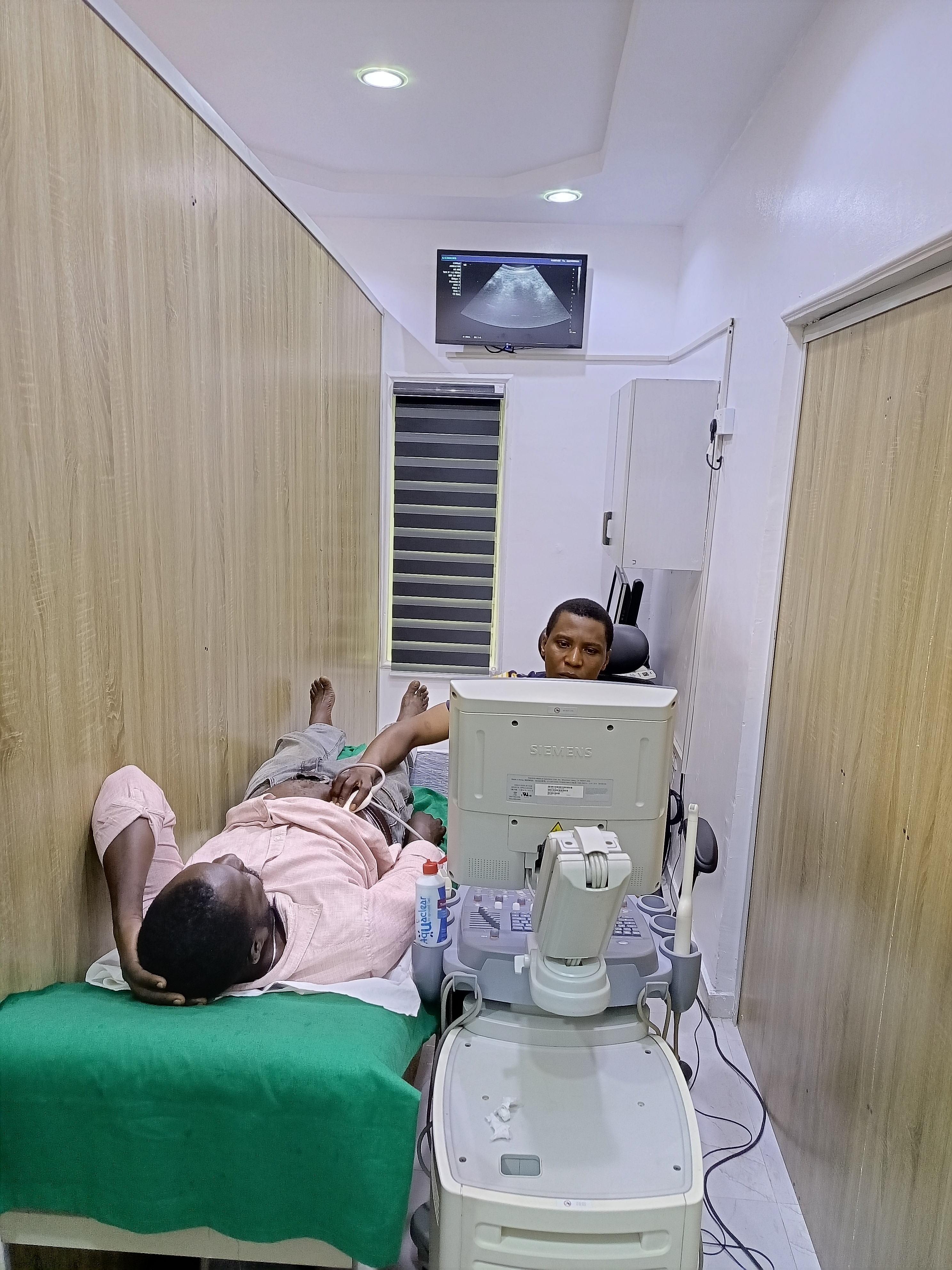
Ultrasound imaging, also known as sonography, is a non-invasive diagnostic technique that uses high-frequency sound waves to produce real-time images of the body’s internal structures. Unlike X-rays or CT scans, ultrasound does not use ionizing radiation, making it a safe option, especially for pregnant women and pediatric patients. Ultrasound is commonly used to visualize soft tissues, organs, blood flow, and even developing fetuses.
Ultrasound is a versatile, safe, and cost-effective imaging technique used in a wide range of medical fields. Its ability to provide real-time, radiation-free images makes it a critical tool in diagnosing, monitoring, and guiding treatments for various conditions. Advances in technology continue to enhance ultrasound’s diagnostic capabilities, ensuring its ongoing relevance in modern healthcare.
An ultrasound machine consists of a transducer (probe) that emits high-frequency sound waves into the body. These sound waves travel through tissues and are reflected back when they hit different structures. The returning echoes are captured by the transducer and are then processed by a computer to create visual images on a monitor.